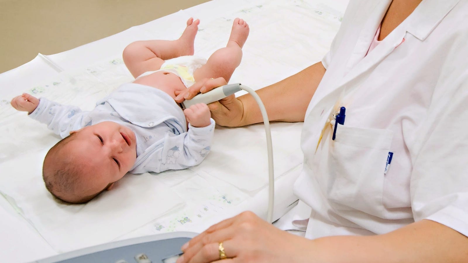
It is standard practice to recommend fetal ultrasound scanning of the fetus for mothers who have a genetic ultrasound scan at 18-20 weeks that shows a cardiac abnormality or a suspected genetic abnormality or who are at high risk for cardiac abnormalities. At our institute, we also perform infant ultrasound scans, which allows us to detect possible heart problems at this age.
Ultrasound scans of the heart, also known as echocardiography, use ultrasound to show the heart on a computer screen. It is also possible to perform a Doppler test, in which the ultrasound machine emits high-frequency waves, some of which are absorbed by the body and some of which are reflected back. This method not only allows simple images of the heart to be taken, but also measures the speed and direction of the blood flow in the heart.
The combination of the two tests can provide the paediatric cardiologist with qualitative information about the anatomical and functional state of the infant’s heart.
What does paediatric cardiology do?
Paediatric cardiology deals with heart disease in children, some of which is congenital and some of which is acquired heart disease. The most common congenital heart diseases are cardiac malformations.
Paediatric cardiology is carried out by a specially trained paediatrician who is skilled in the diagnosis and treatment of congenital and childhood-acquired heart diseases and arrhythmias.
Please note that we can examine children weighing up to 30 kg in our paediatric cardiology clinic.
What are the reasons to see a paediatric cardiologist?
For a screening test if:
- there is a family history of a heart development disorder or heart disease,
- developmental abnormalities of other organs,
- or suspected cardiac malformations in foetal life.
In the event of a complaint or symptom where heart disease or cardiac arrhythmia is suspected. This includes, for example:
- murmurs heard above the heart (heart murmur),
- bluish-purple discoloration of the skin (cyanosis),
- failure to develop weight,
- frequent, recurrent lung disease,
- rapid or strong palpitations, chest pain,
- sudden onset of fatigue,
- reduced exercise capacity.
How is the paediatric cardiology assessment carried out?
The paediatric cardiology examination is made up of several parts. In all cases, children are examined in the presence and with the active participation of their parents, in a relaxed and friendly environment.
The presentation of previous records and a child care record book can help in the diagnosis of the disease.
The paediatric cardiological examination consists of the following tests:
Physical examination, during which the child is palpated, the heart is listened to, and, if necessary, blood pressure and oxygen saturation are measured;
ECG examination, which provides information on the electrical activity of the heart;
cardiac ultrasound scan, which uses special equipment to show the structure and function of the heart in different ways, as a function of heart cycles and time.
Other extended tests: exercise ECG, 24-hour blood pressure monitoring (ABPM), Holter ECG monitoring and laboratory tests may be required based on complaints to ensure an accurate diagnosis.
With all this information, we will summarise the results and provide a detailed and personalised assessment.
Further actions, suggestions and a date for a follow-up examination will be given in the form of a written opinion.


